

Specialist Veterinary Arthroscopy equipment
Stryker 1088 3 chip camera and console
Dynonics 1.9mm, 2.4mm & 2.7mm arthroscopes
Stryker xenon light source
Styker TPS & Shaver
Arthrocare bipolar / RF coblation
Arthrex Continuous Wave III arthroscopy Pump
Stryker SDC 2 image recorder
Shoulder Arthroscopy- Osteochrondrosis OCD

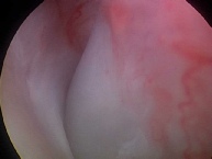
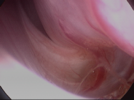
Shoulder arthroscopy- normal dog

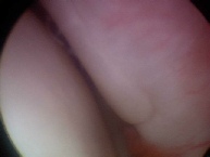
Shoulder arthroscopy- OCD lesion dog

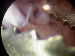
Above- OCD flap
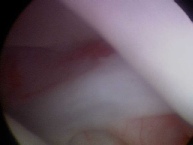
Above- shoulder OCD flap removal with arthroscopic grasping forceps
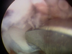
Above- subchondral bone bed after OCD flap removal, before debridement. After removal of the main fragments, the subchondral bone bed is now curretted until healthly bleeding bone is reached, ensuring all cartilage fragments are removed. This now allows the lesion to heal with replacement of the diseased cartilage with a new fibrocartilage surface.

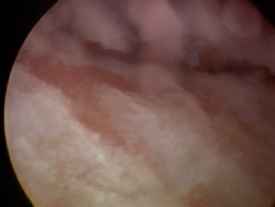
Above- The remaining areas of the joint are inspected for disease and loose flap fragments and flushed.

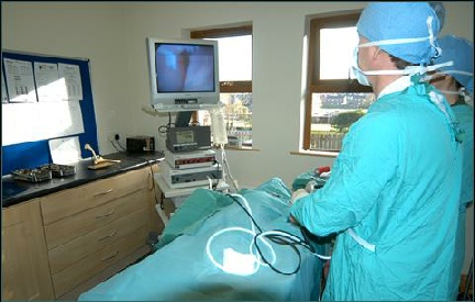
Because arthroscopy is a minimally invasive surgery, both shoulders can be examined and treated arthroscopically at one surgery. Due to the small surgical sites of 3mm diameter there is greatly reduced post operative pain compared to open joint surgery so most dogs are walking on the operated legs immediately after surgery, and recover more quickly that after open surgery.
Effective pain medications are given intra-articularly immediately post op and followed up with injectable and oral medications for several days
| elbow arthroscopy key hole surgery dog ireland |
| elbow dysplasia |
| vet-arthroscopy-shoulder ocd-dog |
| shoulder arthroscopy dog- ligament injury |
| stifle arthroscopy dogs |
| hip arthroscopy dog |
| spinal xray myelogram |
| GSDA A stamp |
| elbow score |
| hip score |
| hip score guide for owners |
| directions |
| site map |