

Specialist Veterinary Arthroscopy equipment
Stryker 1088 3 chip camera and console
Dynonics 1.9mm, 2.4mm & 2.7mm arthroscopes
Stryker xenon light source
Styker TPS & Shaver
Arthrocare bipolar / RF coblation
Arthrex Continuous Wave III arthroscopy Pump
Stryker SDC 2 image recorder
Elbow Dysplasia- FCP, UAP, OCD, Joint Incongruity

Elbow dysplasia is a broad term to cover 4 conditions of the elbow which may occur on their own or in conjunction with some of the others- each condition requires specific treatment
Fragmented coronoid process FCP
Ununited anconeal process- UAP
Joint incongruity
Osteochrondritis dissecans of the medial humeral condyle- OCD

Fragmented coronoid process
Ununited anconeal process UAP
Joint incongruity- this may be one of the most important factors in the development of elbow disease and may lead to the formation of some of the other conditions referred to as elbow dysplasia. It may be seen a step at the level of the ulnar and radial bones as they articulate with the humerus leading to uneven wear of the joint surfaces which may lead to cartilage erosions and may be associated with FCP. This incongruity may be difficult to fully appreciate as it may occur at different levels within the joint, eg radius and ulnar at rest, dynamic incongruity depending on the position of the foot- especially pronation and supination, or between the curved portions of the ulnar and humerus.


Osteochrondritis dissecans of the medial humeral condyle (OCD)
- a cartilage flap may be present on the medial humeral condyle with exposure of the subchrondral bone causing lameness, pain and osteoarthritis. Treatment for small focal flaps is by removal of the flap and debridement of the underlying bed. Small areas will heal by fibrocartilage repair, however larger defects, in particular if incongruity is present, may also benefit from a proximal ulnar osteotomy. Ulnar osteotomy involves cutting the ulnar bone at an oblique angle below the elbow joint and allowing it to heal over the following weeks. This additional treatment results in significant short term lameness while the ulnar heals but may benefit the dog long-term by allowing an improvement in joint incongruity.
Unfortunately if OCD is present, FCP is also frequently present requiring subtotal coronoidectomy.
Ongoing research into cartilage grafting techniques may provide additional benefit in these cases.
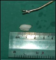
1.7mm x 12mm cartilage flap removed by arthroscopic surgery from an OCD lesion on the medial humeral condyle of a 7 month old chocolate labrador.


FCP relates to tiny crack formations of over small area of the ulnar bone (the coronoid process) which come together leading to fissures or fragments to form. Not all cases have obvious fragments and only have coronoid disease. FCP is best visualised by arthroscopy (key hole examination) to diagnose and assess the entire elbow for co-existing conditions such as kissing lesions on the humerus &OCD. The entire area of affected coronoid may be removed along with any free fragments. For more on FCP
Ununited anconeal process (UAP) occurs because of failure of this small piece of bone on the ulnar to fuse onto the rest of the bone by 20 weeks of age. UAP is seen fairly commonly in German Shepherd dogs and we have found that proximal ulnar ostectomy has lead to a reduction in lameness and a fusion of the UAP in most cases where the UAP is quite stable. This may be assessed arthroscopically. Additionally the ununited anconeal process UAP may be repaired with a lag screw placement into the fragment using a guide wire and a cannulated screw. This is particularly useful if the UAP is unstable. Arthroscopic examination of the elbows is recommended as co-existing FCP may also be present in a percentage of cases.
Various treatments are recommended for the various conditions included under the term “elbow dysplasia” and veterinary opinions vary and alternative methods of treatment are currently undergoing research and clinical trials.
Unfortunately the exposure of the bone underneath the cartilage (subchondral bone) to the joint will stimulate the development of osteoarthritis. No treatment is available which will prevent osteoarthritis developing once it has begun, and all cases will require management of the elbow osteoarthritis by weight control, exercise modulation (regular controlled exercise), NSAIDs (anti inflammatory) medications and pain control.
“Elbow dysplasia” is perhaps not the most useful term as it may lead to confusion. Each of the four conditions are individual conditions and may occur on their own, or co-exist in the same joint. OCD and FCP for example have different underlying reasons for development and genetic factors.
| elbow arthroscopy key hole surgery dog ireland |
| elbow dysplasia |
| vet-arthroscopy-shoulder ocd-dog |
| shoulder arthroscopy dog- ligament injury |
| stifle arthroscopy dogs |
| hip arthroscopy dog |
| spinal xray myelogram |
| GSDA A stamp |
| elbow score |
| hip score |
| hip score guide for owners |
| directions |
| site map |